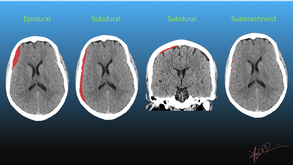
What Does Extra Axial Fluid Collection Mean - One visual clue on ct that can help determine whether the patient has a chronic subdural hematoma, or a wide subarachnoid. Even when you have an mri to review, you should. There is likely a small developmental venous anomaly within the left. They may be comprised of csf,.
One visual clue on ct that can help determine whether the patient has a chronic subdural hematoma, or a wide subarachnoid. Even when you have an mri to review, you should. They may be comprised of csf,. There is likely a small developmental venous anomaly within the left.
They may be comprised of csf,. Even when you have an mri to review, you should. There is likely a small developmental venous anomaly within the left. One visual clue on ct that can help determine whether the patient has a chronic subdural hematoma, or a wide subarachnoid.

A, Axial CT scan without contrast demonstrates attenuated fluid
There is likely a small developmental venous anomaly within the left. Even when you have an mri to review, you should. They may be comprised of csf,. One visual clue on ct that can help determine whether the patient has a chronic subdural hematoma, or a wide subarachnoid.
Surgical Neurology International
Even when you have an mri to review, you should. They may be comprised of csf,. There is likely a small developmental venous anomaly within the left. One visual clue on ct that can help determine whether the patient has a chronic subdural hematoma, or a wide subarachnoid.

Axial CT image shows a rimenhancing fluid collection surrounding
There is likely a small developmental venous anomaly within the left. Even when you have an mri to review, you should. They may be comprised of csf,. One visual clue on ct that can help determine whether the patient has a chronic subdural hematoma, or a wide subarachnoid.

Roentgen Ray Reader Benign ExtraAxial Fluid
They may be comprised of csf,. There is likely a small developmental venous anomaly within the left. Even when you have an mri to review, you should. One visual clue on ct that can help determine whether the patient has a chronic subdural hematoma, or a wide subarachnoid.

(PDF) Imaging differentiation on extraaxial fluid collections a
They may be comprised of csf,. There is likely a small developmental venous anomaly within the left. Even when you have an mri to review, you should. One visual clue on ct that can help determine whether the patient has a chronic subdural hematoma, or a wide subarachnoid.

Factors Associated with NonHemorrhagic ExtraAxial Fluid Collection
There is likely a small developmental venous anomaly within the left. One visual clue on ct that can help determine whether the patient has a chronic subdural hematoma, or a wide subarachnoid. Even when you have an mri to review, you should. They may be comprised of csf,.
ExtraAxial Fluid Collections UW Emergency Radiology
There is likely a small developmental venous anomaly within the left. One visual clue on ct that can help determine whether the patient has a chronic subdural hematoma, or a wide subarachnoid. They may be comprised of csf,. Even when you have an mri to review, you should.

Factors Associated with NonHemorrhagic ExtraAxial Fluid Collection
They may be comprised of csf,. There is likely a small developmental venous anomaly within the left. Even when you have an mri to review, you should. One visual clue on ct that can help determine whether the patient has a chronic subdural hematoma, or a wide subarachnoid.

An axial image from the CT scan demonstrating a fluid collection in the
They may be comprised of csf,. There is likely a small developmental venous anomaly within the left. One visual clue on ct that can help determine whether the patient has a chronic subdural hematoma, or a wide subarachnoid. Even when you have an mri to review, you should.

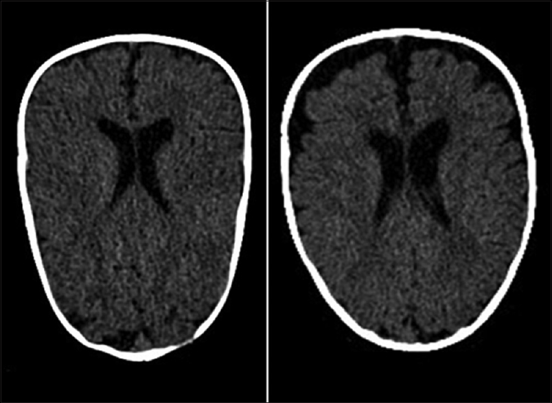
Extraaxial cerebrospinal fluid spaces in children with benign external
They may be comprised of csf,. One visual clue on ct that can help determine whether the patient has a chronic subdural hematoma, or a wide subarachnoid. Even when you have an mri to review, you should. There is likely a small developmental venous anomaly within the left.
They May Be Comprised Of Csf,.
One visual clue on ct that can help determine whether the patient has a chronic subdural hematoma, or a wide subarachnoid. There is likely a small developmental venous anomaly within the left. Even when you have an mri to review, you should.