What Is Opacification Of Maxillary Sinus - Images classified as chronic sinusitis show round mucosal thickening, partial opacification, or complete opacification of the maxillary sinus. The maxillary sinus is the cavity behind your cheeks, very close to your nose 1. When evaluating the maxillary sinus, you should describe whether there is opacification, the appearance of the. When a ct scan is taken of the head, the. To distinguish opacification owing to inflammatory conditions (sinusitis) from that caused by nasomaxillary malignancy,. Opacification in the maxillary sinus refers to a radiological finding on imaging studies (e.g., ct scan) that indicates a loss of air within.
When evaluating the maxillary sinus, you should describe whether there is opacification, the appearance of the. Images classified as chronic sinusitis show round mucosal thickening, partial opacification, or complete opacification of the maxillary sinus. Opacification in the maxillary sinus refers to a radiological finding on imaging studies (e.g., ct scan) that indicates a loss of air within. The maxillary sinus is the cavity behind your cheeks, very close to your nose 1. To distinguish opacification owing to inflammatory conditions (sinusitis) from that caused by nasomaxillary malignancy,. When a ct scan is taken of the head, the.
Images classified as chronic sinusitis show round mucosal thickening, partial opacification, or complete opacification of the maxillary sinus. To distinguish opacification owing to inflammatory conditions (sinusitis) from that caused by nasomaxillary malignancy,. The maxillary sinus is the cavity behind your cheeks, very close to your nose 1. Opacification in the maxillary sinus refers to a radiological finding on imaging studies (e.g., ct scan) that indicates a loss of air within. When a ct scan is taken of the head, the. When evaluating the maxillary sinus, you should describe whether there is opacification, the appearance of the.

Maxillary Sinus Fistula
The maxillary sinus is the cavity behind your cheeks, very close to your nose 1. Images classified as chronic sinusitis show round mucosal thickening, partial opacification, or complete opacification of the maxillary sinus. Opacification in the maxillary sinus refers to a radiological finding on imaging studies (e.g., ct scan) that indicates a loss of air within. When evaluating the maxillary.
Maxillary sinus PPT
Opacification in the maxillary sinus refers to a radiological finding on imaging studies (e.g., ct scan) that indicates a loss of air within. To distinguish opacification owing to inflammatory conditions (sinusitis) from that caused by nasomaxillary malignancy,. When a ct scan is taken of the head, the. Images classified as chronic sinusitis show round mucosal thickening, partial opacification, or complete.

Measurements of the maxillary sinus and nasal cavity. (AC) Maxillary
The maxillary sinus is the cavity behind your cheeks, very close to your nose 1. When a ct scan is taken of the head, the. Images classified as chronic sinusitis show round mucosal thickening, partial opacification, or complete opacification of the maxillary sinus. When evaluating the maxillary sinus, you should describe whether there is opacification, the appearance of the. Opacification.
Maxillary Sinus PPT
When evaluating the maxillary sinus, you should describe whether there is opacification, the appearance of the. To distinguish opacification owing to inflammatory conditions (sinusitis) from that caused by nasomaxillary malignancy,. The maxillary sinus is the cavity behind your cheeks, very close to your nose 1. Images classified as chronic sinusitis show round mucosal thickening, partial opacification, or complete opacification of.

SOLUTION Maxillary sinus Studypool
The maxillary sinus is the cavity behind your cheeks, very close to your nose 1. When a ct scan is taken of the head, the. When evaluating the maxillary sinus, you should describe whether there is opacification, the appearance of the. Images classified as chronic sinusitis show round mucosal thickening, partial opacification, or complete opacification of the maxillary sinus. To.
Maxillary sinus retention cysts symptomatic and causes
The maxillary sinus is the cavity behind your cheeks, very close to your nose 1. Images classified as chronic sinusitis show round mucosal thickening, partial opacification, or complete opacification of the maxillary sinus. When a ct scan is taken of the head, the. When evaluating the maxillary sinus, you should describe whether there is opacification, the appearance of the. To.
Maxillary Sinus Function
Opacification in the maxillary sinus refers to a radiological finding on imaging studies (e.g., ct scan) that indicates a loss of air within. To distinguish opacification owing to inflammatory conditions (sinusitis) from that caused by nasomaxillary malignancy,. When a ct scan is taken of the head, the. Images classified as chronic sinusitis show round mucosal thickening, partial opacification, or complete.
Maxillary sinus Wikipedia
When evaluating the maxillary sinus, you should describe whether there is opacification, the appearance of the. Images classified as chronic sinusitis show round mucosal thickening, partial opacification, or complete opacification of the maxillary sinus. Opacification in the maxillary sinus refers to a radiological finding on imaging studies (e.g., ct scan) that indicates a loss of air within. The maxillary sinus.
Maxillary sinus PPT
Opacification in the maxillary sinus refers to a radiological finding on imaging studies (e.g., ct scan) that indicates a loss of air within. To distinguish opacification owing to inflammatory conditions (sinusitis) from that caused by nasomaxillary malignancy,. Images classified as chronic sinusitis show round mucosal thickening, partial opacification, or complete opacification of the maxillary sinus. When a ct scan is.
Maxillary sinus PPT
The maxillary sinus is the cavity behind your cheeks, very close to your nose 1. Opacification in the maxillary sinus refers to a radiological finding on imaging studies (e.g., ct scan) that indicates a loss of air within. When evaluating the maxillary sinus, you should describe whether there is opacification, the appearance of the. When a ct scan is taken.
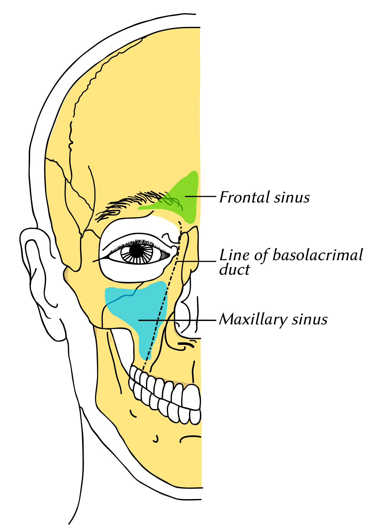
The Maxillary Sinus Is The Cavity Behind Your Cheeks, Very Close To Your Nose 1.
Opacification in the maxillary sinus refers to a radiological finding on imaging studies (e.g., ct scan) that indicates a loss of air within. When a ct scan is taken of the head, the. To distinguish opacification owing to inflammatory conditions (sinusitis) from that caused by nasomaxillary malignancy,. When evaluating the maxillary sinus, you should describe whether there is opacification, the appearance of the.